

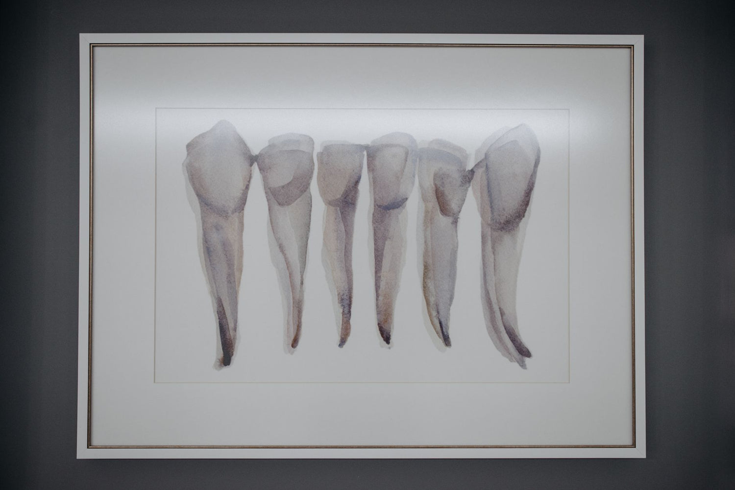
The History of Anatomy Wall Charts: Science Meets Art
Few objects in the history of education are as visually striking and intellectually rich as the anatomy wall chart. These large-format illustrations, pinned to classroom walls, hung in lecture halls, and displayed in physicians' offices, represent a remarkable intersection of scientific discovery and artistic mastery. Long before photography or digital imaging, a skilled illustrator's hand was the only means of transmitting the secrets of the human body across generations. To understand the anatomy wall chart history is to trace one of the most compelling stories in the evolution of both science and visual culture.
Before the Charts: Early Anatomical Illustration

The story begins long before the printing press made mass reproduction possible. In the medieval period, anatomical knowledge was largely inherited from ancient Greek and Roman texts, particularly those of Galen, and illustrations were schematic at best, symbolic diagrams rather than faithful representations of the human form. These early images were more philosophical than practical, designed to convey cosmological ideas about the body rather than to guide a surgeon's hand or a student's understanding.
Everything changed in the early 16th century with the extraordinary work of Leonardo da Vinci. Between approximately 1490 and 1513, Leonardo conducted dozens of human dissections and filled over 200 pages with anatomical drawings of breathtaking precision. He depicted muscles in layers, rendered bones from multiple angles, and illustrated the mechanics of the heart with an understanding that would not be surpassed for centuries. Leonardo's drawings were never published during his lifetime, which means their immediate scientific impact was limited, yet they stand today as perhaps the most beautiful anatomical illustrations ever created, a testament to what happens when a supreme artist turns his eye to the human body.
The true turning point for anatomical illustration as a public, educational medium came with the Flemish physician Andreas Vesalius. In 1543, he published De Humani Corporis Fabrica, a monumental seven-volume work containing extraordinarily detailed woodcut engravings produced in collaboration with artists from Titian's workshop. Vesalius corrected hundreds of errors in Galen's ancient texts, and his illustrations, showing dissected figures posed dramatically against Renaissance landscapes, set the standard for anatomical imagery for the next two centuries. This was the true birth of the history of anatomical illustration as a rigorous, art-driven scientific discipline.
The 17th and 18th Centuries: Refinement and Ambition

Following Vesalius, European anatomists competed fiercely to produce ever more ambitious illustrated atlases. The 17th century saw the rise of copper engraving as the dominant technique, allowing for finer detail and greater tonal range than the woodcuts of the previous century. Works such as Govard Bidloo's Anatomia Humani Corporis (1685), illustrated by the Dutch master Gerard de Lairesse, pushed anatomical illustration into the realm of high art, with dissected specimens arranged on draped tables and set against elaborate backgrounds. The line between scientific document and artistic masterpiece had never been thinner.
In the 18th century, the Scottish anatomist William Hunter and his brother John produced works that combined clinical precision with neoclassical elegance. Meanwhile, the Italian school of wax modeling, centered in Bologna and Florence, created three-dimensional anatomical figures of astonishing realism, further demonstrating the era's obsession with rendering the body in all its complexity. These were luxury objects, however, expensive atlases and museum pieces accessible only to wealthy collectors and elite medical institutions. The idea of a practical, affordable teaching aid that could hang on any classroom wall was still a century away.
What this period established, crucially, was a visual vocabulary for the human body that would inform every anatomy wall chart that followed. The conventions of the écorché figure (a body with skin removed to reveal underlying musculature), the layered dissection view, and the isolated organ study all became standard formats during these two centuries. Artists and anatomists had, together, invented a visual language for the interior of the human body, one that remains recognizable and influential to this day.
The 19th Century: Mass Production and the Classroom Chart

The industrial revolution transformed anatomical illustration from an elite art form into a mass educational medium. The key technology was lithography, invented in 1796 by Alois Senefelder and rapidly refined throughout the early 19th century. Lithography allowed images to be reproduced quickly, cheaply, and in large formats, for the first time, it became economically feasible to print anatomical illustrations large enough to be read from across a room. The anatomy wall chart as we know it was born.
By the mid-19th century, German publishing houses, particularly those based in Leipzig and Stuttgart, had become the world leaders in producing scientific wall charts for educational use. These Wandtafeln (literally "wall tablets") covered not only human anatomy but botany, zoology, geology, and physics. The anatomical charts produced by publishers such as Friedrich Eduard Bilz and later the firm of Lehmann were remarkable objects: printed on heavy paper or linen, often measuring three or four feet in height, and featuring color lithography of extraordinary quality. These charts were designed to teach entire classrooms simultaneously, in an era when textbooks were scarce and projectors did not yet exist.
The production of these charts required a new kind of professional: the scientific illustrator, trained in both anatomy and draftsmanship, who could translate the findings of medical research into clear, accurate, visually compelling images. Many of these artists remain anonymous today, their work absorbed into institutional collections or lost entirely. Yet their charts hung in schools and medical colleges across Europe and North America for decades, shaping the anatomical understanding of generations of students. These vintage anatomy posters were, in a very real sense, the textbooks of their age.
Scientific Accuracy and Artistic Craft: An Evolving Partnership

One of the most fascinating aspects of anatomy wall chart history is the ongoing negotiation between scientific accuracy and artistic convention. A wall chart must be accurate enough to teach correctly, but it must also be legible from a distance, visually organized, and aesthetically engaging enough to hold a student's attention. These are not always the same requirements. Color, for example, was always a compromise: veins are conventionally shown in blue and arteries in red, a convention that has no basis in the actual color of living tissue but that makes the circulatory system dramatically easier to understand at a glance.
Similarly, the practice of showing multiple layers of the body simultaneously, muscles overlaid on bones, organs visible through a transparent torso, is a visual fiction that no surgeon ever encounters in the operating room. Yet it is an enormously effective teaching device, allowing a single image to communicate information that would otherwise require dozens of sequential illustrations. The great anatomy chart artists were masters of this kind of productive simplification, stripping away complexity where necessary while preserving the essential truths of human structure.
As the 19th century gave way to the 20th, the arrival of photography posed an interesting challenge to anatomical illustration. Photographs of cadavers and dissections were certainly more literal than drawings, but they were also messier, harder to read, and less pedagogically effective. Illustrated charts retained their dominance in classrooms well into the mid-20th century, precisely because a skilled artist could do something a camera could not: selectively emphasize what mattered most. This is why the illustrated anatomy chart remained an essential tool even as photography transformed journalism, portraiture, and documentary science.
The Mid-20th Century: Iconic Charts and Enduring Aesthetics

The mid-20th century produced what many collectors and design enthusiasts consider the golden age of the anatomy wall chart. American and European publishers refined their techniques to produce charts that were not only scientifically up-to-date but visually stunning, bold, clean, and graphic in a way that reflected the modernist design sensibility of the postwar era. Companies such as Denoyer-Geppert in the United States and Somso in Germany became household names in educational supply, their charts appearing in virtually every biology classroom and medical school in the Western world.
These mid-century scientific wall charts had a visual confidence that distinguishes them from both their Victorian predecessors and their later, more clinical successors. The color palettes were vivid but harmonious; the typography was bold and purposeful; the compositions were carefully balanced to guide the eye across complex information. Many of these charts are now recognized as significant examples of mid-century graphic design, collected and displayed as art objects entirely independent of their original educational function. Their appeal lies in exactly the combination of qualities that made them effective teaching tools: clarity, beauty, and the sense that behind every line lies genuine knowledge.
It is worth noting that this era also saw the rise of the cool science poster as a cultural object beyond the classroom. Anatomical images appeared in popular magazines, on the covers of science publications, and in the offices of architects, designers, and intellectuals who admired their aesthetic qualities. The anatomy poster had crossed over from educational tool to cultural artifact, a transition that continues to shape how we think about and display these images today. If you are drawn to the aesthetic of scientific illustration, exploring our Science Posters collection offers a wonderful range of options to bring that tradition into your own space.
Vintage Anatomy Posters as Home Decor Today

Today, vintage anatomy posters and their modern reproductions enjoy remarkable popularity as home decor. The reasons are not difficult to understand. These images possess a rare combination of qualities that few decorative objects can match: they are intellectually serious, visually striking, historically resonant, and available in a wide range of styles to suit almost any interior aesthetic. A well-chosen anatomy chart brings depth and character to a space in a way that purely decorative art sometimes cannot.
The appeal is particularly strong among those who work in medicine, biology, or related fields, but it extends far beyond professional circles. Design-conscious homeowners have discovered that antique science posters make compelling focal points in living rooms, home offices, and studies. The combination of aged paper tones, careful hand-lettering, and the intrinsic drama of the human body gives these images a presence that modern digital prints often struggle to replicate. For those who want to complement their anatomical wall art with three-dimensional scientific objects, our human anatomy skeleton model makes an exceptional companion piece, bringing the same spirit of scientific curiosity into sculptural form.
The enduring appeal of the anatomy wall chart also reflects something deeper about our relationship with scientific knowledge. These images remind us that understanding the world, including our own bodies, is one of the most fundamentally human endeavors. They connect us to centuries of physicians, artists, and teachers who believed that knowledge, rendered beautifully, could change how people see themselves and their world. For those who wish to surround themselves with objects that celebrate that tradition, the Biology ambiance collection offers a curated selection of decor inspired by the life sciences in all their visual richness.
Why the Anatomy Wall Chart Still Matters
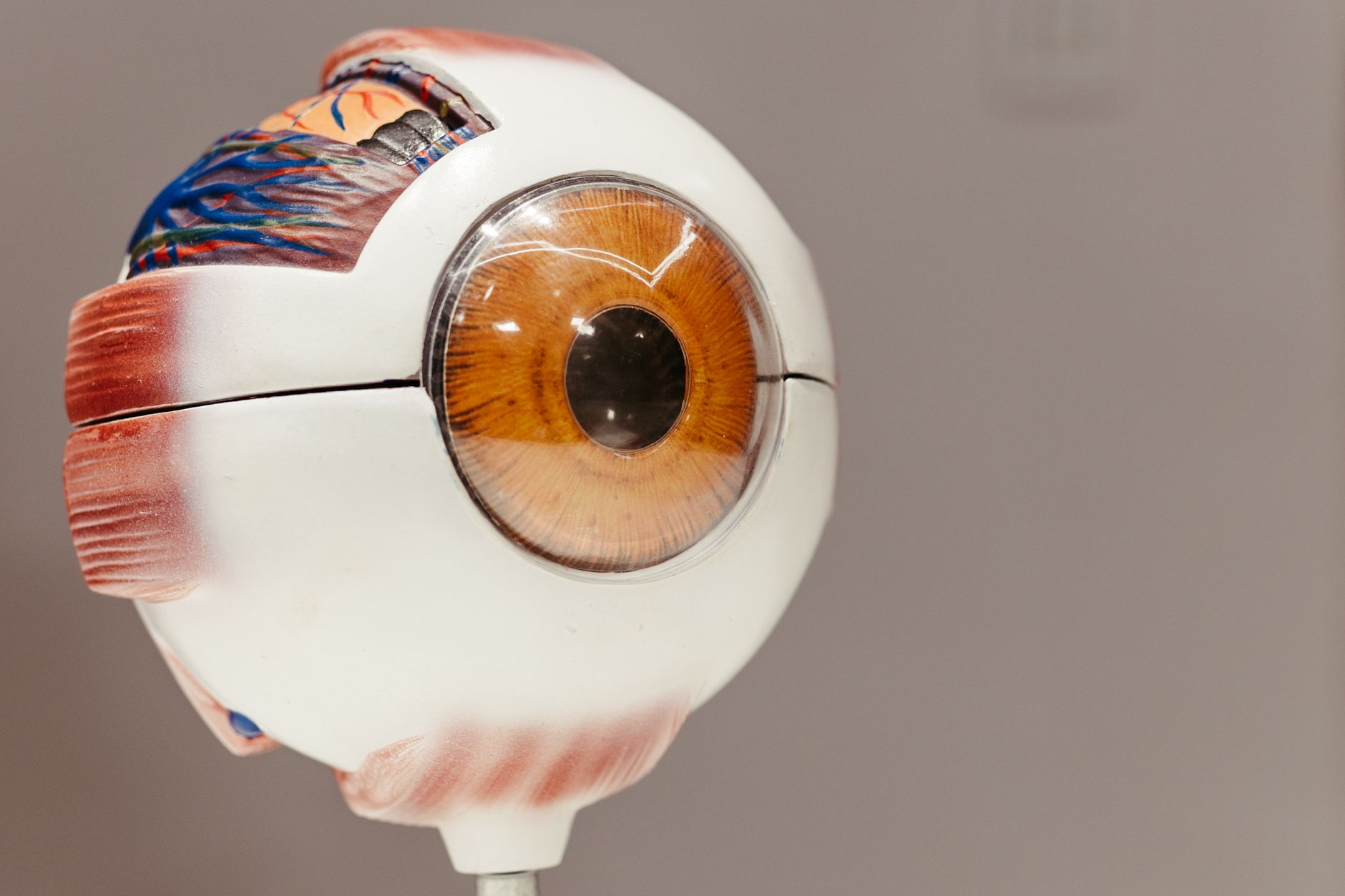
In an age of digital imaging, 3D scanning, and interactive anatomical software, one might ask whether the hand-drawn anatomy chart has any remaining relevance beyond nostalgia. We would argue that it has, and not merely as a decorative object. The illustrated anatomy chart embodies a particular kind of knowledge: synthesized, interpreted, and communicated with the intention of being understood. It is the product of a human mind making decisions about what matters, what to emphasize, and how to make the invisible visible. That process of thoughtful interpretation is something no algorithm has yet replicated.
Furthermore, the history of anatomical illustration is inseparable from the broader history of how humans have understood themselves. Every major advance in anatomical knowledge, from Vesalius's correction of Galen to Harvey's discovery of blood circulation to the mapping of the nervous system, was accompanied by new visual representations that made those advances comprehensible to a wider audience. The anatomy chart was never merely a picture of the body; it was an argument about what the body is and how it works. That argumentative, persuasive quality is part of what makes these images so compelling even centuries after they were created.
As collectors, decorators, and science enthusiasts rediscover these extraordinary objects, they are participating in a tradition that stretches back to Leonardo's notebooks and Vesalius's woodcuts. The anatomy wall chart is one of the great achievements of Western visual culture, a form that demanded the best from both scientists and artists, and that produced, at its finest, images of genuine and lasting beauty. Whether you encounter them in a museum, a medical school, or on the wall of a thoughtfully decorated home, they reward careful attention and repay it with wonder.
The history of anatomy wall charts is, ultimately, the history of human curiosity made visible. From Leonardo's secret notebooks to the lithographed classroom charts of the Victorian era, from the bold mid-century posters that defined a generation of scientific education to the antique science posters now celebrated as decorative art, these images have always been more than illustrations, they are records of the ongoing human effort to understand the body we inhabit. We invite you to explore our collections and bring a piece of that extraordinary history into your own home. Whether you are a lifelong science enthusiast or simply someone who appreciates the rare beauty of knowledge rendered with care and craft, there has never been a better time to celebrate the art of anatomical illustration.



Leave a comment
All comments are moderated before being published.
This site is protected by hCaptcha and the hCaptcha Privacy Policy and Terms of Service apply.